Cobalamin (vitamin B12) is an essential water-soluble cofactor required for two enzymatic reactions in human cells: the cytosolic remethylation of homocysteine to methionine by methionine synthase, and the mitochondrial isomerization of methylmalonyl-coenzyme A (CoA) to succinyl-CoA by methylmalonyl-CoA mutase. Inherited disorders affecting affecting intestinal absorption, plasma transport, cellular uptake, or intracellular processing produce a spectrum of multisystem diseases that present from the neonatal period through adulthood with hematologic, neurologic, psychiatric, ophthalmologic, renal, hepatic, thromboembolic, and cardiovascular manifestations. These conditions are classified by classified according to their molecular genetic defects, historically designated as complementation groups (cblA–cblJ), into groups that affect extracellular handling of cobalamin (intrinsic factor deficiency [GIF], Imerslund-Gräsbeck syndrome [CUB, AMN], transcobalamin deficiency [TCN2], transcobalamin receptor deficiency [CD320]) and groups that affect intracellular processing (including cblA, cblB, cblC, cblD, cblE, cblF, cblG, cblJ and related intracellular cobalamin-processing defects, including the most frequent disorder, cblC disease, caused by biallelic variants in MMACHC). Advances in tandem mass-spectrometry newborn screening, rapid genomic sequencing, and parenteral hydroxocobalamin-based protocols have substantially improved outcomes for early-treated infants, while liver transplantation and emerging gene-addition and genome-editing strategies are reshaping management of isolated methylmalonic acidemia due to methylmalonyl-CoA mutase deficiency. This review summarizes the biochemistry, classification, clinical phenotypes, diagnostic strategy, and current and emerging therapies for inherited disorders of cobalamin metabolism.
Keywords: Vitamin B12; Cobalamin metabolism; Inherited metabolic disorders; Methylmalonic acidemia; cblC disease; MMACHC; Hydroxocobalamin; Newborn screening
Vitamin B12 (cobalamin) was isolated in 1948 from liver extracts as the curative factor for pernicious anemia, representing a landmark discovery in hematology and nutritional medicine. However, the spectrum of inherited disorders resulting from defective absorption, transport, cellular uptake, and intracellular processing of this cobalt-containing corrinoid was not fully elucidated until several decades later through pioneering biochemical and cellular studies, notably those conducted by Rosenblatt, Watkins, and colleagues. These investigations established the framework for the classification of intracellular cobalamin-processing disorders, many of which continue to be designated by their historical complementation group nomenclature (cblA, cblB, cblC, and related defects).
Cobalamin occupies a unique position among vitamins because it serves as the precursor of two metabolically active cofactors: methylcobalamin, required for the cytosolic remethylation of homocysteine to methionine by methionine synthase, and adenosylcobalamin, required for the mitochondrial conversion of methylmalonyl-CoA to succinyl-CoA by methylmalonyl-CoA mutase [1,6]. Defects affecting intestinal absorption, plasma transport, cellular uptake, intracellular trafficking, cofactor synthesis, or enzymatic utilization disrupt one or both of these pathways and generate characteristic biochemical signatures, including methylmalonic acidemia, hyperhomocysteinemia, or their combination. These metabolic profiles frequently provide important diagnostic clues and often permit localization of the underlying defect before molecular confirmation [19,24].
Although individually rare, inherited disorders of cobalamin metabolism collectively constitute an important group of treatable inborn errors of metabolism. Their clinical manifestations are remarkably heterogeneous and may present from the neonatal period through adulthood with hematologic, neurologic, psychiatric, ophthalmologic, renal, thromboembolic, and cardiovascular complications [4,6,21]. Affected patients may initially present with failure to thrive, megaloblastic anemia, developmental delay, neurocognitive deterioration, thrombotic microangiopathy, pulmonary hypertension, or chronic kidney disease, often posing substantial diagnostic challenges across multiple medical specialties.
The clinical significance of these disorders extends beyond their rarity. Advances in tandem mass spectrometry–based newborn screening, molecular diagnostics, and hydroxocobalamin-based treatment protocols have substantially improved outcomes, particularly when therapy is initiated before irreversible organ damage occurs. Early diagnosis has become increasingly important as several inherited cobalamin disorders are now recognized as among the most treatable inborn errors of intermediary metabolism [4,6,21].
Furthermore, major therapeutic innovations are reshaping the management of selected disorders. In particular, liver transplantation has emerged as an option for severe forms of isolated methylmalonic acidemia, while gene-addition, messenger RNA–based, and genome-editing strategies are under active investigation and may transform future therapeutic approaches [6,21].
This review provides an updated overview of the biochemistry, molecular classification, clinical manifestations, focused on neurological and psychiatric manifestations, diagnostic strategies, and current and emerging therapies for inherited disorders of cobalamin metabolism, emphasizing recent advances that continue to improve prognosis and long-term outcomes for affected patients.
Dietary cobalamin is released from food proteins by gastric acid and pepsin and binds to haptocorrin (formerly known as R-binder), which protects the vitamin from degradation within the acidic gastric environment (Figure 1) [13,16]. In the duodenum, pancreatic proteases degrade haptocorrin, allowing cobalamin to bind intrinsic factor, a glycoprotein secreted by gastric parietal cells [13,17]. The intrinsic factor–cobalamin complex resists further proteolysis and is recognized in the terminal ileum by the cubam receptor, a multimeric complex composed of cubilin and amnionless, which mediates receptor-dependent endocytosis into enterocytes [12,17]. Cobalamin is subsequently exported into the portal circulation bound to transcobalamin (TCN2), the plasma transport protein responsible for delivering the vitamin to virtually all nucleated cells through the transcobalamin receptor CD320 [10,11,38].
Following receptor-mediated endocytosis, the transcobalamin–cobalamin complex undergoes lysosomal degradation, and free cobalamin is exported into the cytosol through the coordinated action of LMBRD1 and ABCD4, the proteins defective in the cblF and cblJ disorders, respectively [24,29]. The cytosolic chaperone MMACHC then catalyzes the reductive decyanation or dealkylation of incoming cobalamin species, generating cob(II)alamin, a central intermediate from which the vitamin is directed toward one of two metabolically active cofactor forms. In the cytosol, cobalamin is processed through the methionine synthase pathway involving MTR and its reductase MTRR to generate and maintain methylcobalamin, the cofactor required by methionine synthase for the remethylation of homocysteine to methionine using 5-methyltetrahydrofolate as the methyl donor. Alternatively, cobalamin is targeted to mitochondria, a process involving MMADHC, where ATP(I)alamin adenosyltransferase (ATR, encoded by MMAB) converts it into adenosylcobalamin, the cofactor required by methylmalonyl-CoA mutase (MMUT) for the isomerization of L-methylmalonyl-CoA to succinyl-CoA in the catabolism of branched-chain amino acids, odd-chain fatty acids, and cholesterol side chains [1,6,24].
Most intracellular defects located proximal to this metabolic branch point result in combined methylmalonic acidemia and hyperhomocysteinemia, whereas defects affecting only one downstream pathway produce isolated biochemical phenotypes characterized by either methylmalonic acidemia or disorders of homocysteine remethylation [19,24].
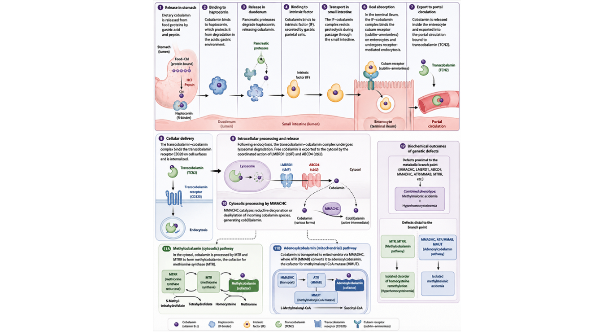
Figure 1: Integrated Gastrointestinal Absorption and Intracellular Metabolism of Cobalamin (Vitamin B12): Transport Proteins, Receptor-Mediated Uptake, and Genetic Disorders.
Figure legend: Schematic representation of cobalamin (vitamin B12) handling from dietary release to intracellular cofactor activation. In the stomach, protein-bound cobalamin is liberated by gastric acid and pepsin and binds haptocorrin (R-binder), which protects the vitamin from acidic degradation. In the duodenum, pancreatic proteases degrade haptocorrin, allowing transfer of cobalamin to intrinsic factor (IF), secreted by gastric parietal cells. The IF–cobalamin complex remains stable through the small intestine and is absorbed in the terminal ileum via the cubam receptor complex (cubilin–amnionless), mediating receptor-dependent endocytosis into enterocytes. Within enterocytes, cobalamin is released into the circulation bound to transcobalamin II (TCN2), which delivers the vitamin to peripheral tissues via the CD320 receptor. Following cellular uptake, the transcobalamin–cobalamin complex is degraded in lysosomes, and free cobalamin is exported into the cytosol through LMBRD1 and ABCD4. Cytosolic processing by MMACHC generates cob(II)alamin, a central intermediate directed toward two major metabolic fates: (1) methylcobalamin synthesis in the cytosol via MTR/MTRR for methionine synthase–dependent remethylation of homocysteine, and (2) mitochondrial conversion to adenosylcobalamin via MMADHC trafficking and MMAB (ATR) activity for methylmalonyl-CoA mutase (MMUT)–dependent conversion of methylmalonyl-CoA to succinyl-CoA. Genetic defects at different levels of this pathway produce distinct biochemical phenotypes. Proximal defects (e.g., MMACHC, LMBRD1, ABCD4, MMADHC, MMAB, MTRR) commonly result in combined methylmalonic acidemia and hyperhomocysteinemia, whereas distal defects in the remethylation or mitochondrial branches lead to isolated homocysteine remethylation disorders or isolated methylmalonic acidemia, respectively.
Inherited disorders of cobalamin (vitamin B12) metabolism are traditionally classified according to the step of the pathway affected. Three major functional levels are recognized: defects of intestinal uptake and absorption, defects of plasma transport, and defects of intracellular cobalamin processing and trafficking. The latter group is further subdivided into historical somatic-cell complementation groups (cblA–cblJ), with additional recently characterized regulatory entities such as cblX and cblK [24,27,29].
Defects of cobalamin absorption include intrinsic factor deficiency, either autoimmune or congenital, and Imerslund–Gräsbeck syndrome. The latter is caused by biallelic pathogenic variants in CUBN or AMN, encoding the cubam receptor complex. This receptor is required for ileal uptake of the intrinsic factor–cobalamin complex. Clinically, these disorders typically present in childhood with megaloblastic anemia. Proteinuria is frequently associated, reflecting the role of cubilin in proximal renal tubular protein reabsorption [12,17].
Transcobalamin deficiency, caused by biallelic variants in TCN2, impairs cellular delivery of absorbed cobalamin despite normal or near-normal serum total vitamin B12 concentrations. This discrepancy is explained by the predominance of circulating cobalamin bound to haptocorrin rather than transcobalamin. The disease usually presents in early infancy with failure to thrive, diarrhea, pancytopenia, megaloblastic anemia, and recurrent infections. It is highly responsive to pharmacologic parenteral hydroxocobalamin therapy [21-23]. Fewer than 50 genetically confirmed cases have been reported worldwide, underscoring the need for clinical suspicion even in the context of apparently normal serum cobalamin levels [22].
The most frequent inherited defect of intracellular cobalamin metabolism is the cblC complementation group, caused by biallelic pathogenic variants in MMACHC. Its estimated incidence ranges from 1:100,000 to 1:200,000 live births, depending on the population [6,8].
Early-onset cblC disease, accounting for approximately 90% of cases, presents within the first months of life with a severe multisystem phenotype. Clinical features include hypotonia, feeding difficulties, failure to thrive, megaloblastic anemia, progressive microcephaly, seizures, and neuro-ophthalmologic involvement, including maculopathy. Renal complications have increasingly been recognized, including thrombotic microangiopathy resembling atypical hemolytic uremic syndrome, particularly in association with specific variants such as c.80A>G [2,4,5,7,34].
Late-onset cblC disease may present from childhood to adulthood. The phenotype is more heterogeneous and often neurologic, including subacute combined degeneration of the spinal cord, cognitive decline, psychiatric manifestations (depression or psychosis), and thromboembolic events. Diagnosis is frequently delayed in this form due to its variable and nonspecific presentation [2,3,25].
Defects of the cblA and cblB groups, caused by variants in MMAA and MMAB, respectively, impair adenosylcobalamin synthesis. They typically present with isolated methylmalonic aciduria without hyperhomocysteinemia. Clinically, cblA disease is generally more responsive to hydroxocobalamin therapy than cblB disease, a distinction with prognostic and therapeutic relevance [18,20,35].
The cblD complementation group, caused by variants in MMADHC, is phenotypically heterogeneous. Depending on the affected protein domain, it may result in combined methylmalonic aciduria and homocystinuria (cblD-MMA/HC), isolated methylmalonic aciduria (cblD-MMA), or isolated homocystinuria (cblD-HC) [17,19].
Defects of the cblE and cblG groups, due to pathogenic variants in MTRR and MTR, respectively, impair methionine synthase function. They result in isolated homocystinuria with low or low-normal methionine levels. This biochemical profile distinguishes them from classical homocystinuria due to cystathionine β-synthase deficiency, in which methionine is elevated [19,24].
Finally, the cblF and cblJ groups, caused by variants in LMBRD1 and ABCD4, impair lysosomal export of cobalamin following receptor-mediated endocytosis. These disorders lead to combined methylmalonic aciduria and homocystinuria and are characterized by intralysosomal accumulation of unmetabolized cobalamin [24,29].
Inherited disorders of cobalamin metabolism constitute a clinically and genetically heterogeneous group of diseases affecting sequential steps of absorption, transport, and intracellular processing. Despite their rarity, they are increasingly recognized due to expanded biochemical screening and molecular diagnostics. Early identification is crucial, as several forms—particularly defects of intracellular processing—are responsive to pharmacologic hydroxocobalamin and amenable to improved neurological and systemic outcomes.
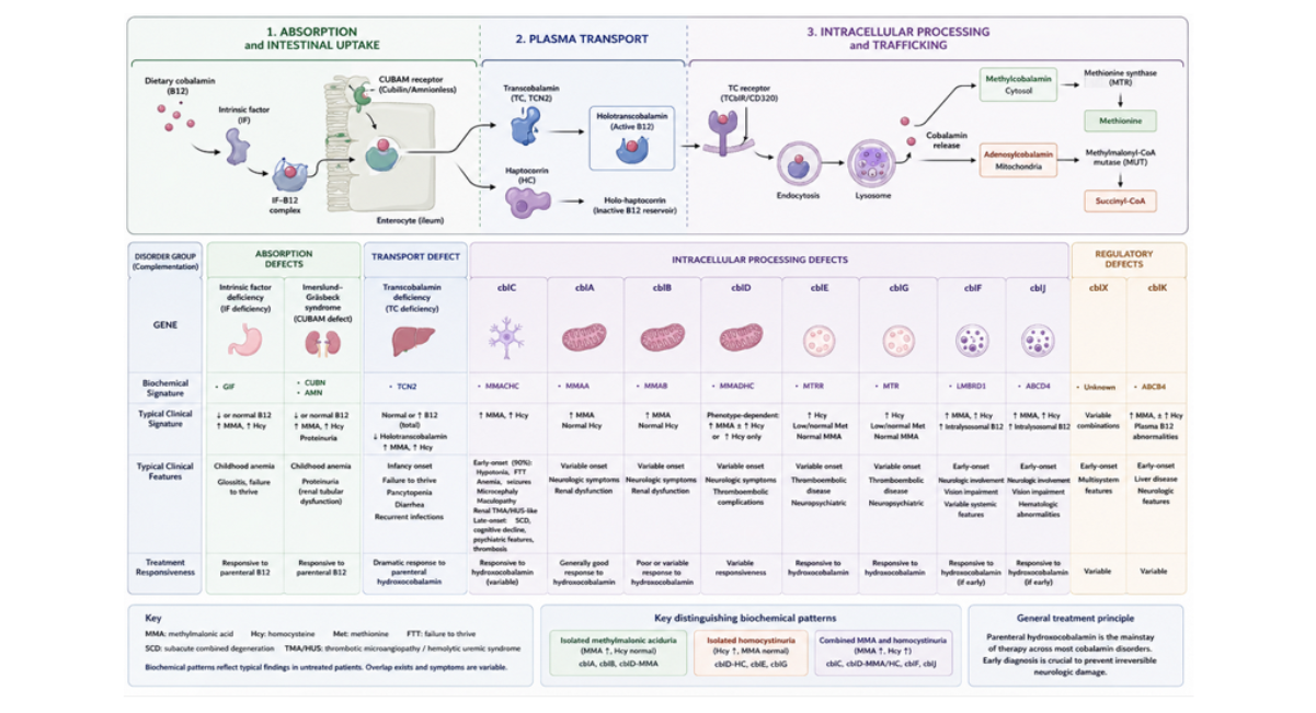
Figure 2: Inherited Disorders of Cobalamin Metabolism: Classification, Genes, Biochemistry and Key Clinical Manifestations.
Inherited disorders of cobalamin metabolism are multisystem diseases characterized by a broad clinical spectrum involving hematologic, neurologic, psychiatric, ophthalmologic, renal, hepatic, thromboembolic, and cardiovascular systems (Table 1). The relative prominence of each organ involvement depends on the age at onset and the underlying biochemical defect, with combined remethylation disorders (particularly cblC) showing the widest systemic expression.
Hematologic involvement is common, particularly in early-onset disease and in disorders affecting methylcobalamin-dependent methionine synthase activity. The typical presentation includes megaloblastic anemia with macrocytosis, hypersegmented neutrophils, and variable pancytopenia. These abnormalities reflect impaired thymidylate and purine synthesis due to functional folate trapping within the one-carbon metabolism pathway [21,33].
Neurologic disease is a major determinant of morbidity. Early-onset forms present with hypotonia, developmental delay, feeding difficulties, and seizures. In later-onset disease, the spectrum includes subacute combined degeneration of the spinal cord, peripheral neuropathy, motor dysfunction, and progressive cognitive decline. Pathophysiology is multifactorial, involving impaired methylation reactions, accumulation of toxic metabolites (methylmalonic acid and homocysteine), mitochondrial dysfunction, and oxidative stress [2,3,29].
Psychiatric involvement is increasingly recognized, particularly in late-onset cblC and related remethylation disorders. Clinical features include behavioral changes, mood disorders (notably depression), cognitive deterioration, psychosis, and, occasionally, acute confusional states. These manifestations may precede overt neurologic signs and contribute to frequent misdiagnosis. Disruption of methylation-dependent neurotransmitter pathways and subcortical white matter dysfunction are thought to play central roles [2,3,25].
Ophthalmologic involvement is a characteristic feature of cblC disease. It typically presents as a pigmentary retinopathy or maculopathy with progressive visual impairment. Retinal changes may be detected early, including in presymptomatic infants identified through newborn screening programs [2,4].
Renal disease ranges from chronic tubulointerstitial nephropathy to acute thrombotic microangiopathy resembling atypical hemolytic uremic syndrome. These complications are most prominent in cblC disease and are attributed to endothelial dysfunction driven by hyperhomocysteinemia, oxidative stress, and microvascular injury [5,34].
Hepatic involvement is less frequent but may occur in severe early-onset forms, particularly cblC disease. Reported abnormalities include hepatomegaly, elevated transaminases, cholestasis, and, in rare cases, acute liver dysfunction. These manifestations likely reflect systemic metabolic toxicity and mitochondrial dysfunction.
Thromboembolic events are a recognized complication, especially in remethylation disorders with elevated homocysteine. Clinical presentations include venous and arterial thrombosis, cerebrovascular events, and microangiopathic processes. Endothelial dysfunction, impaired nitric oxide signaling, and prothrombotic changes contribute to the increased vascular risk.
Cardiovascular involvement is primarily mediated through endothelial dysfunction and thrombotic propensity. Reported manifestations include cardiomyopathy in severe early-onset disease, pulmonary hypertension in selected cases, and vascular complications related to systemic hyperhomocysteinemia. These features are most consistently observed in cblC disease and related remethylation defects [2,3,21,33].
|
Organ system |
Typical clinical manifestations |
Predominant biochemical context |
Key mechanisms |
Most associated disorders |
|
Hematologic |
Megaloblastic anemia, macrocytosis, hypersegmented neutrophils, pancytopenia |
Impaired methylcobalamin / methionine synthase pathway |
Functional folate trapping → impaired thymidylate & purine synthesis |
cblC, cblE, cblG, cblD-HC |
|
Neurologic |
Hypotonia, developmental delay, seizures (early onset); subacute combined degeneration, neuropathy, cognitive decline (late onset) |
Combined MMA + HC or remethylation defects |
Impaired methylation, MMA/Hcy toxicity, mitochondrial dysfunction, oxidative stress |
cblC, cblD, cblE, cblG, cblA/B (MMA phenotype) |
|
Psychiatric |
Depression, psychosis, behavioral changes, cognitive deterioration, acute confusional states |
Mainly remethylation disorders |
Disrupted monoamine metabolism, white matter dysfunction, methylation deficit |
cblC (late-onset), cblE, cblG |
|
Ophthalmologic |
Pigmentary maculopathy, progressive visual loss, retinal dystrophy |
Combined MMA + HC |
Retinal mitochondrial dysfunction, toxic metabolite accumulation |
cblC (hallmark), cblD |
|
Renal |
Tubulointerstitial nephropathy, proteinuria, thrombotic microangiopathy (TMA/HUS-like) |
Mainly cblC |
Endothelial injury, hyperhomocysteinemia, oxidative stress |
cblC (± cblD) |
|
Hepatic |
Hepatomegaly, transaminase elevation, cholestasis, rare acute liver failure |
Severe early-onset multisystem disease |
Mitochondrial dysfunction, systemic metabolic toxicity |
cblC (severe neonatal forms) |
|
Thromboembolic |
Venous thrombosis, arterial thrombosis, stroke, microangiopathy |
Hyperhomocysteinemia (remethylation defects) |
Endothelial dysfunction, prothrombotic state, impaired NO signaling |
cblC, cblE, cblG, cblD |
|
Cardiovascular |
Cardiomyopathy (rare), pulmonary hypertension, vascular complications |
Severe systemic metabolic derangement |
Endothelial dysfunction, homocysteine-mediated vascular injury |
cblC (severe forms), remethylation defects |
Table 1: Systemic clinical manifestations of inherited cobalamin disorders.
Diagnosis begins with measurement of plasma total homocysteine and urinary or plasma methylmalonic acid, which together help localize the underlying defect. Isolated methylmalonic aciduria without hyperhomocysteinemia suggests impairment of adenosylcobalamin synthesis or of methylmalonyl-CoA mutase itself (mut⁰, mut⁻, cblA, cblB), whereas isolated hyperhomocysteinemia without methylmalonic aciduria indicates a defect in methylcobalamin synthesis or methionine synthase function (cblE, cblG) or methylenetetrahydrofolate reductase deficiency. Combined elevation of methylmalonic acid and homocysteine suggests defects affecting both cobalamin-dependent pathways, most typically cblC (MMACHC), cblD (MMA/HC subtype), cblF, cblJ, or disorders of cobalamin absorption and intracellular transport [19,24,30].
Serum total cobalamin levels are frequently normal in inherited intracellular cobalamin processing disorders, as circulating concentrations do not reflect intracellular utilization, which may lead to diagnostic confusion. In this context, holotranscobalamin—the biologically active fraction bound to transcobalamin—may provide a more sensitive functional marker in selected cases [10,38].
Definitive diagnosis requires molecular genetic testing, increasingly performed using multigene panels or exome-based approaches covering MMACHC, MMAA, MMAB, MMADHC, MTRR, MTR, LMBRD1, ABCD4, MMUT, TCN2, CUBN, AMN, and MTHFR, reflecting the substantial phenotypic overlap across complementation groups [19,24,36].
Tandem mass spectrometry-based newborn screening, based on elevated propionylcarnitine (C3) and an increased C3/C2 acylcarnitine ratio, identifies most infants with methylmalonic acidemia, including cblC disease, before symptom onset [36,37,41]. Comparative cohort studies have shown substantially lower mortality and improved neurodevelopmental outcomes in infants diagnosed through newborn screening and treated pre-symptomatically compared with those diagnosed after clinical presentation, supporting the inclusion of these disorders in expanded newborn screening programs [37,41].
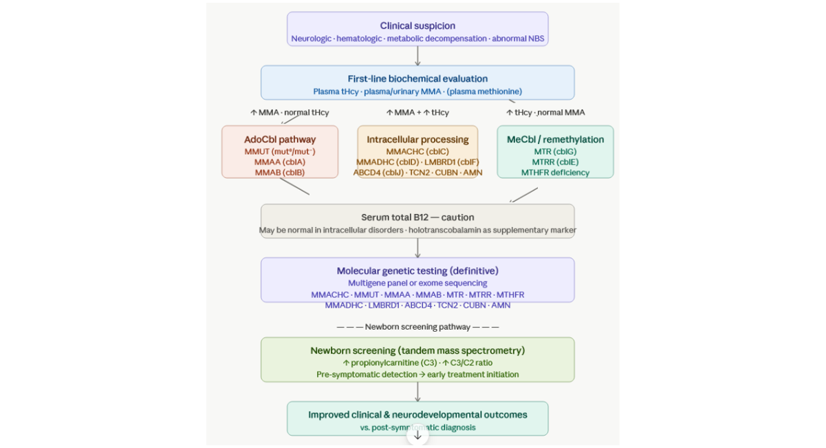
Figure 3: Diagnostic algorithm for inborn errors of cobalamin metabolism.
Figure legend: Clinical suspicion arises from neurologic, hematologic (e.g., megaloblastic anemia), metabolic presentations, and/or abnormal newborn screening (↑ C3 and C3/C2 ratio). Initial biochemical testing includes plasma total homocysteine (tHcy) and methylmalonic acid (MMA). Isolated MMA elevation indicates defects in adenosylcobalamin synthesis or methylmalonyl-CoA mutase (MMUT, MMAA/cblA, MMAB/cblB). Isolated tHcy elevation suggests remethylation defects (MTR/cblG, MTRR/cblE, MTHFR deficiency). Combined MMA and tHcy elevation indicates intracellular cobalamin processing or transport disorders, most commonly MMACHC (cblC), as well as MMADHC (cblD), LMBRD1 (cblF), ABCD4 (cblJ), or transport defects (TCN2, CUBN, AMN). Serum vitamin B12 may be normal; holotranscobalamin may provide additional functional information. Definitive diagnosis relies on multigene panel or exome sequencing targeting the above genes. Newborn screening by tandem mass spectrometry (↑ C3, ↑ C3/C2) enables presymptomatic diagnosis and improved outcomes.
For cobalamin-responsive inborn errors of intracellular cobalamin metabolism, parenteral hydroxocobalamin is the treatment of choice over cyanocobalamin, owing to superior plasma retention, cellular uptake, and intracellular conversion to adenosylcobalamin and methylcobalamin [19,22]. In cblC, cblF, and cblJ disease, treatment typically consists of daily intramuscular or subcutaneous hydroxocobalamin (up to 1 mg/day during acute decompensation), followed by individualized maintenance regimens (several times weekly to daily), guided by biochemical response, including plasma total homocysteine and methylmalonic acid [6,19].
Adjunctive therapy includes betaine to promote remethylation of homocysteine via the betaine–homocysteine methyltransferase pathway, together with folinic acid (vitamin B9). Carnitine supplementation is used in cases of secondary carnitine depletion or significant organic acid excretion. Dietary protein restriction is not routinely indicated in cblC, cblF, or cblJ disorders but may be considered in selected patients with methylmalonic acidemia phenotypes and persistent metabolic instability despite optimized medical therapy [6,19].
Transcobalamin deficiency and intrinsic factor–related malabsorption disorders are highly responsive to parenteral cobalamin replacement, with rapid hematologic recovery and variable neurologic outcomes depending on treatment initiation prior to irreversible injury [21-23].
Isolated methylmalonic acidemia due to methylmalonyl-CoA mutase deficiency (mut0/mut–) is generally cobalamin-unresponsive and managed with protein restriction, carnitine supplementation, and aggressive prevention of catabolic stress. By contrast, cblA disease is typically cobalamin-responsive and associated with a more favorable metabolic and clinical outcome under hydroxocobalamin therapy [18,20,35].
In severe, recurrent metabolic decompensation refractory to optimal medical management, liver transplantation reduces systemic methylmalonic acid burden but does not normalize metabolite production, while improving survival and reducing frequency of metabolic crises. However, renal and neurologic complications may persist after transplantation [25,27,28,32,33] Kidney transplantation has been used in selected patients with advanced renal disease, providing partial metabolic correction through restoration of enzymatic activity in renal tissue [29].
Emerging therapies include adeno-associated viral vector–mediated gene addition, lipid nanoparticle–delivered mRNA encoding methylmalonyl-CoA mutase, and CRISPR-based genome editing strategies. Preclinical studies in animal models suggest a selective growth advantage for corrected hepatocytes, supporting the feasibility of durable metabolic correction without solid-organ transplantation [30,31].
In adults, inherited disorders of intracellular cobalamin metabolism, particularly cblC disease and related complementation groups, should be considered in the differential diagnosis of unexplained neurologic syndromes, thromboembolic disease, renal impairment, and atypical hematologic abnormalities, even in the presence of normal serum vitamin B12 concentrations [6,15,19]. The diagnostic key is the measurement of plasma total homocysteine, often markedly elevated, frequently accompanied by increased methylmalonic acid, reflecting combined remethylation and adenosylcobalamin pathway dysfunction [6,38]. Use of serum holotranscobalamin level determination may be of interest. Recognition is critical because prompt parenteral hydroxocobalamin administration, rather than cyanocobalamin, is associated with superior intracellular availability and metabolic correction, particularly in cblC, cblF, and cblJ disorders [19,22]. Adjunctive therapy with betaine enhances remethylation of homocysteine via the betaine–homocysteine methyltransferase pathway, while folinic acid and carnitine supplementation may be indicated in selected patients depending on metabolic profile and organ involvement [19]. Although classical methylmalonyl-CoA mutase deficiency (mut0/mut−) is typically unresponsive to cobalamin, cblA disease often retains responsiveness and may exhibit substantial biochemical and clinical improvement under hydroxocobalamin therapy [18,20]. Delayed diagnosis in adults may result in irreversible neurologic injury, chronic kidney disease, or recurrent vascular events; however, treatment can still reduce metabolic burden and stabilize disease progression. In severe or refractory cases, liver or combined liver–kidney transplantation reduces systemic metabolite accumulation but does not fully normalize biochemical abnormalities, while emerging gene-based and mRNA therapies offer potential future disease-modifying strategies supported by preclinical models demonstrating selective advantage of corrected hepatocytes [25,29-31].
Inherited disorders of cobalamin metabolism span a continuum from highly treatable conditions responsive to parenteral hydroxocobalamin to forms requiring lifelong dietary management, organ transplantation, or emerging genomic therapies. Because clinical presentations are protean and serum cobalamin concentrations are frequently normal or only mildly reduced, diagnosis depends on a high index of suspicion supported by targeted biochemical testing — plasma homocysteine and methylmalonic acid — followed by confirmatory genetic testing. Expanded newborn screening has measurably improved outcomes by enabling presymptomatic treatment, and ongoing development of gene-based therapies for cobalamin-unresponsive methylmalonic acidemia promises to further transform the natural history of these historically devastating but increasingly manageable disorders [36,37,40].
Conflicts of interest: The authors declare no competing interests.
Acknowledgments: The authors thank the patients, their families, and the clinicians and researchers of the CARE B12 group for their contributions to the advancement of knowledge in inherited disorders of cobalamin metabolism. We also acknowledge the contribution of E. Andrès, J.E. Terrade, and N. Lorenzo-Villalba for their scientific input and support in the development of this work.
Author Contributions: E. Andrès conceived the work, supervised the overall manuscript development, and provided critical revision for important intellectual content. J.E. Terrade and N. Lorenzo-Villalba contributed to literature review, data synthesis, and drafting of the manuscript. All authors contributed to the interpretation of the literature, critically revised the manuscript for important intellectual content, and approved the final version for submission.
Data sources and tools: Literature review and manuscript preparation were informed by structured searches of PubMed and Google Scholar. Reference management was performed using EndNote. Artificial intelligence–assisted drafting and language refinement were performed using ChatGPT and Claude (Anthropic) as supportive tools; all scientific content was verified and curated by the authors.

 Download PDF